Addressing Mortons Neuroma

Overview

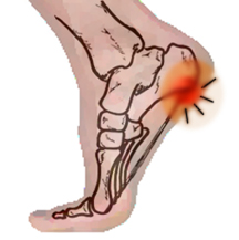
Morton’s neuroma, also called Morton’s metatarsalgia, Morton’s disease, Morton’s neuralgia, Morton metatarsalgia, Morton nerve entrapment, plantar neuroma, or intermetatarsal neuroma is a benign (non-cancerous) growth of nerve tissue (neuroma) that develops in the foot, usually between the third and fourth toes (an intermetatarsal plantar nerve, most commonly of the third and fourth intermetatarsal spaces). It is a common, painful condition.

Causes

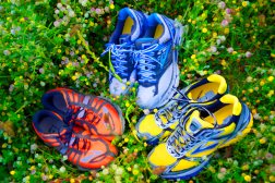
Although the exact cause for this condition is unclear, a number of factors can contribute to the formation of a neuroma. Biomechanical deformities, such as a high-arched foot or a flat foot, can lead to the formation of a neuroma. These foot types bring on instability around the toe joints, leading to the development of the condition. Trauma can cause damage to the nerve, resulting in inflammation or swelling of the nerve. Improper footwear that causes the toes to be squeezed together is problematic. Avoid high-heeled shoes higher than two inches. Shoes at this height can increase pressure on the forefoot area. Repeated stress, common to many occupations, can create or aggravate a neuroma.

Symptoms

The most common presenting complaints include pain and dysesthesias in the forefoot and corresponding toes adjacent to the neuroma. Pain is described as sharp and burning, and it may be associated with cramping. Numbness often is observed in the toes adjacent to the neuroma and seems to occur along with episodes of pain. Pain typically is intermittent, as episodes often occur for minutes to hours at a time and have long intervals (ie, weeks to months) between a single or small group of multiple attacks. Some patients describe the sensation as “walking on a marble.” Massage of the affected area offers significant relief. Narrow tight high-heeled shoes aggravate the symptoms. Night pain is reported but is rare.

Diagnosis

The diagnosis of interdigital neuroma is usually made by physical examination and review of the patient’s medical history.MRI ad High Definition Ultrasound examination may be useful to confirm the diagnoses however they may still not be 100% reliable. The commonest reason for this is de to natural substances present in between the metatarsal heads and between the fat pad and the intermetatarsal ligament. These natural substances i.e. bursa, fat, capsular thickening and even bony growths, can all be a factor in the impingement process and may need to be surgically cleared.

Non Surgical Treatment

The good news is that the pain can often be relieved fairly easily with the right softer styled orthotic (even in those cases where there is concurrent plantar plate tears and capsulitis!), but its important to remeber that even if your no longer in pain, there is no magic cure to speeding up the healing process so one must take care of their feet for 6-12 weeks. As a rule of thumb a neuroma should always be treated conservatively where possible. This means icing and resting the area, trying to remove the causative factors, and providing postural control and support via metatarsal domes or, if needed, specialised pre-made or custom made orthotics.plantar neuroma

Surgical Treatment

Surgery to remove the neuroma may be recommended if more conservative treatment does not solve the problem. While surgery usually relieves or completely removes the symptoms, it often leaves a permanent numb feeling at the site of the neuroma.


Hammertoe Repair Without Surgery

HammertoeOverview
A Hammertoes is a term used to describe a crooked, deviated, or contracted toe. Although the condition usually stems from muscle imbalance, it is often aggravated by poor-fitting shoes or socks that cramp the toes. Over a period of years, the tendons that move the toe up and down begin to pull the toe with unequal tension, and the toe then begins to buckle or become contracted, causing an abnormal “v”-shaped bending of the little toes. Patients with this condition often experience pain, swelling, redness and stiffness in the affected toes.

Causes
It is possible to be born with a hammer toe, however many people develop the deformity later in life. Common causes include tightened tendons that cause the toe to curl downward. Nerve injuries or problems with the spinal cord. Stubbing, jamming or breaking a toe. Having a stroke. Being a diabetic. Having a second toe that is longer than the big toe. Wearing high heels or tight shoes that crowd the toes and don?t allow them to lie flat. Aging.

HammertoeSymptoms
The most obvious sign of hammertoes are bent toes, other symptoms may include pain and stiffness during movement of the toe. Painful corns on the tops of the toe or toes from rubbing against the top of the shoe’s toe box. Painful calluses on the bottoms of the toe or toes. Pain on the bottom of the ball of the foot. Redness and swelling at the joints.

Diagnosis
Hammer toes may be easily detected through observation. The malformation of the person’s toes begin as mild distortions, yet may worsen over time – especially if the factors causing the hammer toes are not eased or removed. If the condition is paid attention to early enough, the person’s toes may not be permanently damaged and may be treated without having to receive surgical intervention. If the person’s toes remain untreated for too long, however the muscles within the toes might stiffen even more and will require invasive procedures to correct the deformity.

Non Surgical Treatment
Apply a commercial, nonmedicated hammertoe pad around the bony prominence of the hammertoe. This will decrease pressure on the area. Wear a shoe with a deep toe box. If the hammertoe becomes inflamed and painful, apply ice packs several times a day to reduce swelling. Avoid heels more than two inches tall. A loose-fitting pair of shoes can also help protect the foot while reducing pressure on the affected toe, making walking a little easier until a visit to your podiatrist can be arranged. It is important to remember that, while this treatment will make the hammertoe feel better, it does not cure the condition. A trip to the podiatric physician?s office will be necessary to repair the toe to allow for normal foot function. Avoid wearing shoes that are too tight or narrow. Children should have their shoes properly fitted on a regular basis, as their feet can often outgrow their shoes rapidly. See your podiatric physician if pain persists.

Surgical Treatment
Probably the most frequent procedure performed is one called a Post or an Arthroplasty. In this case a small piece of bone is removed from the joint to straighten the toe. The toe is shortened somewhat, but there is still motion within the toe post-operatively. In other cases, an Arthrodesis is performed. This involves fusing the abnormally-contracted joint. The Taylor procedure fuses only the first joint in the toe, whereas the Lambrinudi procedure fuses both joints within the toe. Toes which have had these procedures are usually perfectly straight, but they take longer to heal and don’t bend afterwards. A Hibbs procedure is a transfer of the toe’s long extensor tendon to the top of the metatarsal bone. The idea of this procedure is to remove the deforming cause of the hammertoes (in this case, extensor substitution), but to preserve the tendon’s function in dorsifexing the foot by reattaching it to the metatarsals. Fortunately, the Gotch (or Gotch and Kreuz) procedure–the removal of the base of the toe where it attaches to the foot, is done less frequently than in years past. The problem with this procedure is that it doesn’t address the problem at the level of the deformity, and it causes the toe to become destabilized, often resulting in a toe that has contracted up and back onto the top of the foot. You can even have an Implant Arthroplasty procedure, where a small, false joint is inserted into place. There are several other procedures, as well.


Over-Pronation Of The Foot Cause And Treatment

Overview

Pronation is a turning outward of the foot at the ankle, which allows the foot to flatten. The pronation helps to absorb some of the compressive shock forces, torque conversion, adjustment to uneven ground contours, and maintenance of balance. It is necessary to have a certain amount of pronation to disseminate the energy of the foot-strike. If there is too little or too much pronation injuries may occur. When a foot and ankle pronates to a great degree, we call it over-pronation. Normally you can see the Achilles Tendon run straight down the leg into the heel. If the foot is over-pronated, the tendon will run straight down the leg, but when it lies on the heel it will twist outward. This makes the inner ankle bone much more prominent than the outer ankle bone.Over-Pronation

Causes

Over-pronation is very prominent in people who have flexible, flat feet. The framework of the foot begins to collapse, causing the foot to flatten and adding stress to other parts of the foot. As a result, over-pronation, often leads to Plantar Fasciitis, Heel Spurs, Metatarsalgia, Post-tib Tendonitis and/or Bunions. There are many causes of flat feet. Obesity, pregnancy or repetitive pounding on a hard surface can weaken the arch leading to over-pronation. Often people with flat feet do not experience discomfort immediately, and some never suffer from any discomfort at all. However, when symptoms develop and become painful, walking becomes awkward and causes increased strain on the feet and calves.

Symptoms

If you overpronate, your symptoms may include discomfort in the arch and sole of foot, your foot may appear to turn outward at the ankle, your shoes wear down faster on the medial (inner) side of your shoes. Pain in ankle, shins, knees, or hips, especially when walking or running are classic symptoms of overpronation. Overpronation can lead to additional problems with your feet, ankles, and knees. Runners in particular find that overpronation can lead to shin splints, tarsal tunnel syndrome, plantar fasciitis, compartment syndrome, achilles tendonitis, bunions or hallux valgus, patello-femoral pain syndrome, heel spurs, metatarsalgia.

Diagnosis

People who overpronate have flat feet or collapsed arches. You can tell whether you overpronate by wetting your feet and standing on a dry, flat surface. If your footprint looks complete, you probably overpronate. Another way to determine whether you have this condition is to simply look at your feet when you stand. If there is no arch on the innermost part of your sole, and it touches the floor, you likely overpronate. The only way to truly know for sure, however, is to be properly diagnosed by a foot and ankle specialist.Over-Pronation

Non Surgical Treatment

No matter what the cause in your case, over pronation can be remedied in several ways. Those who are overweight should consider permanently losing weight to naturally alleviate pressure on the ligaments and heel of the foot. Also, you should consult a podiatrist to examine your posture and movement habits. You may be reinjuring yourself due to poor alignment without even knowing it. If you also have lower back problems, this could be a sign of over pronation as a result of misalignment.

Prevention

With every step we take, we place at least half of our body weight on each foot (as we walk faster, or run, we can exert more than twice our body weight on each foot). As this amount of weight is applied to each foot there is a significant shock passed on to our body. Custom-made orthotics will absorb some of this shock, helping to protect our feet, ankles, knees, hips, and lower back.


Calcaneal Apophysitis Facts And Figures

Overview

Children with Sever’s disease, which is also called calcaneal apophysitis, develop microfractures where the Achilles tendon inserts on the calcaneus, the large bone that makes up the heel of the foot. These microfractures cause pain, which can vary depending on the type of activity your child is doing, and is generally worse after activity and improves with rest. Sever’s disease is more common in boys and typically occurs when a child is between 8 and 13 years old. Although it can affect both heels, it more commonly just affects one foot.

Causes

Inflammation occurs at the insertion of the achilles tendon into the back of the heel due to a number of reasons. One or several of the following may cause the initiation of Sever?s disease. Rapid growth spurt. Tight calf muscles. Change in footwear (soccer boots / athletic shoes no heel). Excessive rolling in of feet. Poor warm up routine. Remember this condition usually settles as the growth plate fuses within 6-12 months.

Symptoms

Pain is usually related to activity levels. In most cases the posterior aspect of the calcaneus will be tender. Checking both the medial and lateral aspects of the posterior portion of the growth plate will often show tenderness. Occasionally, the plantar aspect may be tender or both of these locations may be found to be tender. Frequently the Achilles tendon is tight and there may have been a recent increase in activity. The factors contributing to this disorder are similar to those causing plantar fasciitis, but a tight Achilles tendon appears to be a greater contributor than pronation.

Diagnosis

A Podiatrist can easily evaluate your child?s foot, lower limbs and muscular flexibility, to identify if a problem exists. If a problem is identified, a simple treatment plan is put in place. Initial treatment may involve using temporary padding and strapping to control motion or to cushion the painful area and based on the success of this treatment, a long-term treatment plan will be put in place. This long-term treatment plan may or may not involve Foot Supports, Heel Raises, muscle stretching and or strengthening.

Non Surgical Treatment

Treatment depends on the severity of the condition, but may include relative rest and modified activity, a physiotherapist can help work out what, and how much, activity to undertake. Cold packs, apply ice or cold packs to the back of the heels for around 15 minutes after any physical activity, including walking. Shoe inserts, small heel inserts worn inside the shoes can take some of the traction pressure off the Achilles tendons. This will only be required in the short term. Medication, pain-relieving medication may help in extreme cases, but should always be combined with other treatment and following consultation with your doctor). Anti-inflammatory creams are also an effective management tool. Splinting or casting, in severe cases, it may be necessary to immobilise the lower leg using a splint or cast, but this is rare. Time, generally the pain will ease in one to two weeks, although there may be flare-ups from time to time. Correction of any biomechanical issues, a physiotherapist can identify and discuss any biomechanical issues that may cause or worsen the condition. Education on how to self-manage the symptoms and flare-ups of Sever?s disease is an essential part of the treatment.


What Can Cause Heel Pain And Discomfort

Overview

Feet Pain

Heel pain is a very common foot problem. The sufferer usually feels pain either under the heel (planter fasciitis) or just behind it (Achilles tendinitis), where the Achilles tendon connects to the heel bone. Even though Heel Pain can be severe and sometimes disabling, it is rarely a health threat. Heel pain is typically mild and usually disappears on its own, however, in some cases the pain may persist and become chronic (long-term). There are 26 bones in the human foot, of which the heel (calcaneus) is the largest. The human heel is designed to provide a rigid support for the weight of the body. When we are walking or running it absorbs the impact of the foot when it hits the ground, and springs us forward into our next stride. Experts say that the stress placed on a foot when walking may be 1.25 times our body weight, and 2.75 times when running. Consequently, the heel is vulnerable to damage, and ultimately pain.

Causes

There are many possible causes of heel pain. Most commonly it is a chronic, long-term pain that results of some type of faulty biomechanics (abnormalities in the way you walk) that place too much stress on the heel bone and the soft tissues that attach to it. Chronic pain is a common result of standing or walking too many hours in the course of a day, working on concrete, being overweight, wearing poorly-constructed shoes, having an overly-pronated foot type (where the arch collapses excessively) or the opposite–having too high an arch. Women seem to get this slightly more often than men, and while any age can be affected, it usually occurs between 30 and 50 years of age. The other type of heel pain is the sort you get from an acute injury–a bruise to the bone or soft tissue strain resulting from a strenuous activity, like walking, running, or jumping, or from some degree of trauma. While there are dozens of possible causes to heel pain, I will review some of the more common causes. Arch Pain/Plantar Fasciitis. One of those often-painful soft tissue that attaches to heel spurs at the bottom of the foot is called “plantar fascia”. Fascia, located throughout the body, is a fibrous connective tissue similar to a ligament. You can see fascia as some of that white, connective tissue attaching to bones, when you pull apart meat. The “plantar” fascia in our bodies is that fascia which is seen on the bottom (or plantar portion) of the foot, extending from the heel bone to the ball of the foot. Compared to other fascia around the body, plantar fascia is very thick and very strong. It has to be strong because of the tremendous amount of force it must endure when you walk, run or jump. But while the plantar fascia is a strong structure, it can still get injured, most commonly when it is stretched beyond its normal length over long periods of time. Plantar Fascitis. When plantar fascia is injured, the condition is called “plantar fasciitis”, pronounced “plan-tar fash-I-tis”. (Adding “-itis” to the end of a word means that structure is inflamed.) It is sometimes known more simply as ‘fasciitis’. Plantar fasciitis is the most common type of arch pain. Symptoms of plantar fasciitis may occur anywhere along the arch, but it is most common near its attachment to the heel bone.

Symptoms

See your doctor immediately if you have Severe pain and swelling near your heel. Inability to bend your foot downward, rise on your toes or walk normally. Heel pain with fever, numbness or tingling in your heel. Severe heel pain immediately after an injury. Schedule an office visit if you have. Heel pain that continues when you’re not walking or standing. Heel pain that lasts more than a few weeks, even after you’ve tried rest, ice and other home treatments.

Diagnosis

Depending on the condition, the cause of heel pain is diagnosed using a number of tests, including medical history, physical examination, including examination of joints and muscles of the foot and leg, X-rays.

Non Surgical Treatment

Home care, in cases that are not severe, home care is probably enough to get rid of heel pain. Rest, avoid running or standing for long periods, or walking on hard surfaces. Avoid activities that may stress the heels. Ice, place an ice-pack on the affected area for about 15 minutes. Do not place bare ice directly onto skin. Footwear. proper-fitting shoes that provide good support are crucial. Athletes should be particularly fussy about the shoes they use when practicing or competing – sports shoes need to be replaced at specific intervals (ask your trainer). Foot supports, wedges and heel cups can help relieve symptoms.

Surgical Treatment

It is rare to need an operation for heel pain. It would only be offered if all simpler treatments have failed and, in particular, you are a reasonable weight for your height and the stresses on your heel cannot be improved by modifying your activities or footwear. The aim of an operation is to release part of the plantar fascia from the heel bone and reduce the tension in it. Many surgeons would also explore and free the small nerves on the inner side of your heel as these are sometimes trapped by bands of tight tissue. This sort of surgery can be done through a cut about 3cm long on the inner side of your heel. Recently there has been a lot of interest in doing the operation by keyhole surgery, but this has not yet been proven to be effective and safe. Most people who have an operation are better afterwards, but it can take months to get the benefit of the operation and the wound can take a while to heal fully. Tingling or numbness on the side of the heel may occur after operation.

Prevention

Feet Pain

A variety of steps can be taken to avoid heel pain and accompanying afflictions. Wear shoes that fit well-front, back, and sides-and have shock-absorbent soles, rigid shanks, and supportive heel counters. Wear the proper shoes for each activity. Do not wear shoes with excessive wear on heels or soles. Prepare properly before exercising. Warm up and do stretching exercises before and after running. Pace yourself when you participate in athletic activities. Don?t underestimate your body’s need for rest and good nutrition. If obese, lose weight.


Achilles Tendinitis Cause And Treatment

Overview

Achilles TendonAchilles tendinitis is an inflammation (swelling) of the tendon, which usually occurs as a result of overuse injury. Basketball players are the most susceptible to Achilles tendinitis because of the frequent jumping. Any activity requiring a constant pushing off the foot, such as running or dancing, may result in swelling of the tendon.

Causes

Achilles tendinitis may be caused by intensive hill running, sprinting, or stair climbing. Overuse resulting from the natural lack of flexibility in the calf muscles. Rapidly increasing intensity of exercise, especially after a period of inactivity. Sudden and hard contraction of the calf muscles when exerting extra effort, like that in a final sprint or high jump.

Symptoms

There will be a gradual onset of achilles tendon pain over a period of weeks, or even months. The pain will come on during exercise and is constant throughout the training session. Pain will be felt in the achilles tendon when walking especially up hill or up stairs. This is because the achilles is having to stretch further than normal. There is likely to be stiffness in the Achilles tendon especially in the morning or after a long period of rest. This is thought to be due to adhesions between the tendon sheath and the tendon itself. Nodules or lumps may be found in the achilles tendon, particularly 2-4cm above the heel and the skin will appear red. Pain and tenderness will be felt when pressing in on the achilles tendon which is likely to appear thickened or swollen. A creaking sensation may be felt when press the fingers into the sides of the tendon and moving the ankle.This is known as crepitus.

Diagnosis

A podiatrist can usually make the diagnosis by clinical history and physical examination alone. Pain with touching or stretching the tendon is typical. There may also be a visible swelling to the tendon. The patient frequently has difficulty plantarflexing (pushing down the ball of the foot and toes, like one would press on a gas pedal), particularly against resistance. In most cases X-rays don’t show much, as they tend to show bone more than soft tissues. But X-rays may show associated degeneration of the heel bone that is common with Achilles Tendon problems. For example, heel spurs, calcification within the tendon, avulsion fractures, periostitis (a bruising of the outer covering of the bone) may all be seen on X-ray. In cases where we are uncertain as to the extent of the damage to the tendon, though, an MRI scan may be necessary, which images the soft tissues better than X-rays. When the tendon is simply inflamed and not severely damaged, the problem may or may not be visible on MRI. It depends upon the severity of the condition.

Nonsurgical Treatment

Achilles tendinitis can typically be treated at home by following the R.I.C.E. treatment method. Rest. Rest the tendon by avoiding activities that irritate the tendon or increase swelling. However, this does not mean you should be completely inactive for long periods of time, as this can cause stiffness in your joints. It?s still important to stretch in order to maintain strength and flexibility and partake in activities that don?t put direct pressure on the tendon, such as bicycling. Ice. Apply ice to the affected area for 20-minutes at a time, every couple hours, as needed, to reduce swelling and pain. Compression. Use compression bandages to help reduce swelling. Elevation. Elevate your ankle above the level of your heart to help reduce swelling. It is particularly important to do this at night while you sleep. Simply place a pillow or two under your ankle to keep it elevated. Once the tendon has healed, be sure to gradually return to more strenuous activities. If flattened arches contributed to the injury, wear shoes with appropriate support or inserts to prevent the condition from progressing or recurring. If these non-surgical treatments have not been able to provide relief of symptoms after several months, surgery may be performed to remove inflamed tissue. However, this is not usually recommended unless all other options have been exhausted. Consult your doctor for more information about surgical treatment options.

Achilles Tendon

Surgical Treatment

In most surgeries, damaged tissue is cleaned out before surgeons make the necessary repairs. However, a new minimally-invasive surgery to repair a torn Achilles tendon actually uses the damaged tissue to help repair the tear. The percutaneous Achilles repair system, or PARS technique, enables surgeons to better repair a torn Achilles tendon through a smaller incision. This procedure was recently performed at Houston Methodist Hospital to treat an NFL cornerback, getting him back on field for this season.

Prevention

A 2014 study looked at the effect of using foot orthotics on the Achilles tendon. The researchers found that running with foot orthotics resulted in a significant decrease in Achilles tendon load compared to running without orthotics. This study indicates that foot orthoses may act to reduce the incidence of chronic Achilles tendon pathologies in runners by reducing stress on the Achilles tendon1. Orthotics seem to reduce load on the Achilles tendon by reducing excessive pronation,


What Is Plantar Fasciitis And The Right Way To Treat It

Plantar Fascia

Overview

Plantar fasciitis is the most common cause of heel pain. The plantar fascia camera.gif is the flat band of tissue (ligament) that connects your heel bone to your toes. It supports the arch of your foot. If you strain your plantar fascia, it gets weak, swollen, and irritated (inflamed). Then your heel or the bottom of your foot hurts when you stand or walk. Plantar fasciitis is common in middle-aged people. It also occurs in younger people who are on their feet a lot, like athletes or soldiers. It can happen in one foot or both feet.


Causes

When some people stand/walk/run/jump their own anatomy in their ankle joint is not ‘sturdy’ enough to cope with the needed stabilisation of their ankle joint when they are weight bearing. So, their ankle rotates to find a point of stability. By the shin twisting in and the ankle rotating downwards to the inside (along with your body weight, the power of some muscles, and of course, gravity) a huge amount of stress is applied to the plantar fascia until it is stressed beyond it’s normal limits and it starts to ‘tighten up’. It is this tightening up of the plantar fascia under this stress that causes the damage that in turn leads to pain…eventually.


Symptoms

Pain tends to start gradually, often just in the heel, but it can sometimes be felt along the whole of the plantar fascia. The symptoms are initially worse in the morning and mostly after, rather than during, activity. As the condition becomes worse, the symptoms become more persistent.


Diagnosis

Your doctor will check your feet and watch you stand and walk. He or she will also ask questions about your past health, including what illnesses or injuries you have had. Your symptoms, such as where the pain is and what time of day your foot hurts most. How active you are and what types of physical activity you do. Your doctor may take an X-ray of your foot if he or she suspects a problem with the bones of your foot, such as a stress fracture.


Non Surgical Treatment

Orthotics are corrective foot devices. They are not the same as soft, spongy, rubber footbeds, gel heel cups etc. Gel and rubber footbeds may cushion the heels and feet, but they do not provide any biomechanical correction. In fact, gel can do the opposite and make an incorrect walking pattern even more unstable! Orthotic insoles work by supporting the arches while re-aligning the ankles and lower legs. Most people’s arches look quite normal when sitting or even standing. However, when putting weight on the foot the arches lower, placing added tension on the plantar fascia, leading to inflammation at the heel bone. Orthotics support the arches, which reduces the tension and overwork of the plantar fascia, allowing the inflamed tissue to heal. Orthotics needn’t be expensive, custom-made devices. A comprehensive Heel Pain study by the American Orthopaedic Foot and Ankle Society found that by wearing standard orthotics and doing a number of daily exercises, 95% of patients experienced substantial, lasting relief from their heel pain symptoms.

Plantar Fascia


Surgical Treatment

If treatment hasn’t worked and you still have painful symptoms after a year, your GP may refer you to either an orthopaedic surgeon, a surgeon who specialises in surgery that involves bones, muscles and joints, a podiatric surgeon, a podiatrist who specialises in foot surgery. Surgery is sometimes recommended for professional athletes and other sportspeople whose heel pain is adversely affecting their career. Plantar release surgery. Plantar release surgery is the most widely used type of surgery for heel pain. The surgeon will cut the fascia to release it from your heel bone and reduce the tension in your plantar fascia. This should reduce any inflammation and relieve your painful symptoms. Surgery can be performed either as, open surgery, where the section of the plantar fascia is released by making a cut into your heel, endoscopic or minimal incision surgery – where a smaller incision is made and special instruments are inserted through the incision to gain access to the plantar fascia. Endoscopic or minimal incision surgery has a quicker recovery time, so you will be able to walk normally much sooner (almost immediately), compared with two to three weeks for open surgery. A disadvantage of endoscopic surgery is that it requires both a specially trained surgical team and specialised equipment, so you may have to wait longer for treatment than if you were to choose open surgery. Endoscopic surgery also carries a higher risk of damaging nearby nerves, which could result in symptoms such as numbness, tingling or some loss of movement in your foot. As with all surgery, plantar release carries the risk of causing complications such as infection, nerve damage and a worsening of your symptoms after surgery (although this is rare). You should discuss the advantages and disadvantages of both techniques with your surgical team. Extracorporeal shockwave therapy (EST) is a fairly new type of non-invasive treatment. Non-invasive means it does not involve making cuts into your body. EST involves using a device to deliver high-energy soundwaves into your heel. The soundwaves can sometimes cause pain, so a local anaesthetic may be used to numb your heel. It is claimed that EST works in two ways. It is thought to, have a “numbing” effect on the nerves that transmit pain signals to your brain, help stimulate and speed up the healing process. However, these claims have not yet been definitively proven. The National Institute for Health and Care Excellence (NICE) has issued guidance about the use of EST for treating plantar fasciitis. NICE states there are no concerns over the safety of EST, but there are uncertainties about how effective the procedure is for treating heel pain. Some studies have reported that EST is more effective than surgery and other non-surgical treatments, while other studies found the procedure to be no better than a placebo (sham treatment).


Stretching Exercises

Calf stretch. Lean forward against a wall with one knee straight and the heel on the ground. Place the other leg in front, with the knee bent. To stretch the calf muscles and the heel cord, push your hips toward the wall in a controlled fashion. Hold the position for 10 seconds and relax. Repeat this exercise 20 times for each foot. A strong pull in the calf should be felt during the stretch. Plantar fascia stretch. This stretch is performed in the seated position. Cross your affected foot over the knee of your other leg. Grasp the toes of your painful foot and slowly pull them toward you in a controlled fashion. If it is difficult to reach your foot, wrap a towel around your big toe to help pull your toes toward you. Place your other hand along the plantar fascia. The fascia should feel like a tight band along the bottom of your foot when stretched. Hold the stretch for 10 seconds. Repeat it 20 times for each foot. This exercise is best done in the morning before standing or walking.


What Leads To Painful Heel And How To Overcome It

Foot Pain

Overview

A common condition that affects people of all ages. Symptoms include heel pain that is worse upon arising in the morning or standing after prolonged sitting. The pain is caused by inflammation of the plantar fascia, the ligament that connects the heel bone to the toes.


Causes

There are multiple potential causes and contributing factors to plantar fasciitis heel pain. The structure of a person’s foot and the way that they walk or run usually play a significant role in the development of plantar fasciitis. Those with an arch that is lower or higher than the average person are more likely to be afflicted. Overexertion and/or participating in activities that a person is not accustomed to also place a person at risk. This can include a heavy workout, a job change, or even an extended shopping trip. Additionally, inappropriate shoes are also often a factor. Exercising in shoes that are worn out or don’t have enough support and/or wearing inexpensive, flimsy or flat-soled dress or casual shoes are common culprits. In warm climates, such as here in Southern California, people who wear flip-flop sandals or even go barefoot throughout the year increase their chances of developing heel pain. Many athletes and weekend warriors develop heel or arch pain from over-exertion during running or other sports. People who work at jobs that involve long periods of standing, such as grocery checkers, cashiers, warehouse workers, postal workers, and teachers are more susceptible as well. Adults of all ages can develop plantar fasciitis. Heel pain in children is usually caused by a different type of condition.


Symptoms

Symptoms of the plantar fasciitis include a gradual onset of pain under the heel which may radiate into the foot. Tenderness is usually felt under and on the inside of the heel which is initially worse first in the morning but eases as the foot warms up only to return later in the day or after exercise. Stretching the plantar fascia may be painful.


Diagnosis

Your doctor can usually diagnose plantar fasciitis just by talking to you and examining your feet. Rarely, tests are needed if the diagnosis is uncertain or to rule out other possible causes of heel pain. These can include X-rays of the heel or an ultrasound scan of the fascia. An ultrasound scan usually shows thickening and swelling of the fascia in plantar fasciitis.


Non Surgical Treatment

Most people who have plantar fasciitis recover with conservative treatments in just a few months. Pain relievers such as ibuprofen (Advil, Motrin IB, others) and naproxen (Aleve) may ease the pain and inflammation associated with plantar fasciitis. Stretching and strengthening exercises or use of specialized devices may provide symptom relief. These include physical therapy. A physical therapist can instruct you in a series of exercises to stretch the plantar fascia and Achilles tendon and to strengthen lower leg muscles, which stabilize your ankle and heel. A therapist may also teach you to apply athletic taping to support the bottom of your foot. Night splints. Your physical therapist or doctor may recommend wearing a splint that stretches your calf and the arch of your foot while you sleep. This holds the plantar fascia and Achilles tendon in a lengthened position overnight and facilitates stretching. Orthotics. Your doctor may prescribe off-the-shelf heel cups, cushions or custom-fitted arch supports (orthotics) to help distribute pressure to your feet more evenly.

Plantar Fascitis


Surgical Treatment

In cases that do not respond to any conservative treatment, surgical release of the plantar fascia may be considered. Plantar fasciotomy may be performed using open, endoscopic or radiofrequency lesioning techniques. Overall, the success rate of surgical release is 70 to 90 percent in patients with plantar fasciitis. Potential risk factors include flattening of the longitudinal arch and heel hypoesthesia as well as the potential complications associated with rupture of the plantar fascia and complications related to anesthesia.


Symptoms Of Capsulitis of the Second Toe

Pain across the bottom of the foot at any point between the heel and the ball of the foot is often referred to as “arch pain” Although this description is non-specific, most arch pain is due to strain or inflammation Claw Toe of the plantar fascia (a long ligament on the bottom of the foot). Wearing inappropriate footwear or foot problems like athlete’s foot and Morton’s neuroma are some of the factors that cause burning feet sensation.

Orthotics are shoe insoles, custom-made to guide the foot into corrected biomechanics. Orthotics are commonly prescribed to help with hammer toes, heel spurs, metatarsal problems, bunions, diabetic ulcerations and numerous other problems. They also help to minimize shin splints, back pain and strain on joints and ligaments. Orthotics help foot problems by ensuring proper foot mechanics and taking pressure off the parts of your foot that you are placing too much stress on. Dr. Cherine’s mission is to help you realize your greatest potential and live your life to its fullest.

If you see just a thin line connecting the ball of your foot to your heel, you have high arches. If you have flat feet or high arches, you’re more likely to get plantar fasciitis, an inflammation of the tissue along the bottom of your foot. Without proper arch support, you can have pain in your heels, arch, and leg. You can also develop bunions and hammertoes, which can become painful,” says Marlene Reid, a podiatrist, or foot and ankle doctor, in Naperville, IL. Shoes with good arch support and a slightly raised heel can help ward off trouble. Laces, buckles, or straps are best for high arches. See a foot doctor to get fitted with custom inserts for your shoes. Good running shoes, for example, can prevent heel pain, stress fractures , and other foot problems that can be brought on by running. A 2-inch heel is less damaging than a 4-inch heel. If you have flat feet, opt for chunky heels instead of skinny ones, Reid says.Plantar Fasciitis,Pes Planus,Mallet Toe,High Arched Feet,Heel Spur,Heel Pain,Hammer Toe,Hallux Valgus,Foot Pain,Foot Hard Skin,Foot Conditions,Foot Callous,Flat Feet,Fallen Arches,Diabetic Foot,Contracted Toe,Claw Toe,Bunions Hard Skin,Bunions Callous,Bunion Pain,Ball Of Foot Pain,Back Pain

Another solution is to wear custom foot orthotics, like ezWalker ® Performance Custom Orthotics, in your shoes to help correct your body posture, stabilize your balance, relieve pain during follow-through and evenly redistribute your weight on your feet. EzWalker® Custom Orthotics are specifically made for each of your feet to properly support your arches while reducing pressure on the balls of your feet. With ezWalker® Custom Orthotics, you’ll walk from lateral heel to medial forefoot for better biomechanics of your entire body. This condition manifests as a skin lesion that assumes a ring-like pattern. It can affect any region of the body, right from the scalp to the foot. One such common home remedy is the use of bleach. Many people claim that this is a very effective ringworm treatment.Plantar Fasciitis,Pes Planus,Mallet Toe,High Arched Feet,Heel Spur,Heel Pain,Hammer Toe,Hallux Valgus,Foot Pain,Foot Hard Skin,Foot Conditions,Foot Callous,Flat Feet,Fallen Arches,Diabetic Foot,Contracted Toe,Claw Toe,Bunions Hard Skin,Bunions Callous,Bunion Pain,Ball Of Foot Pain,Back Pain

During the average lifetime our feet cover over 70,000 miles, the equivalent of walking four times around the world., so it’s not surprising that problems can occur. Indeed around three-quarters of all adults will experience some sort of problem with their feet at some time. And without treatment most foot complaints will become gradually worse with time. This means people often endure painful conditions for far too long, and the problem can get worse. People often assume nothing can be done to help their condition, but in fact these conditions are extremely treatable. Swollen lump on big toe joint; lump may become numb but also make walking painful.


Identification And Some Useful Solutions

Take a good look at the questions you circled. This method is not foolproof but it is a great way to see all the potential causes or root origins of your back pain. You should see the things you need to improve in order to help you live free of back pain. To ensure long lasting relief from pain, the patient would need to work together as a team with his physicians to treat lower back pain. The treatment for lower back pain may also involve regular home-based exercises, muscle development activities, lifestyle modification, learning the safest way to lift objects and maintaining a healthy body weight.

Arthritis – patients with osteoarthritis commonly experience problems with the joints in the hips, lower back, knees and hands. In some cases spinal stenosis can develop – the space around the spinal cord narrows. Abnormal curvature of the spine – if the spine curves in an unusual way the patient is more likely to experience back pain. An example is scoliosis , when the spine curves to the side. Cancer of the spine – a tumor located on the spine may press against a nerve, resulting in back pain. A large number of patients opt for complementary therapies, as well as conventional treatments; some opt just for complementary therapies.

To reduce pain nurses can encourage patients to bed rest and modification of the position is determined to improve lumbar flexion. Patients are taught to control and adjust the pains that go through the respiratory diaphragm and relaxation can help reduce muscle tension that contributes to lower back pain. Distract patients from pain with other activities such as reading books, watching TV and with imagination. Get plenty of rest. Elevating your feet is also good for your back How can you treat back pain during pregnancy? Use a licensed health care professional such as a chiropractor or massage therapist When to contact your health care provider?back pain during period

Some back pain can be “self-treated”. For example, by applying hot or cold packs, using over-the-counter medications, and even by improving posture. However, a physician should be consulted if pain persists, gets worse, or if arm or leg numbness or weakness, or loss of bowel or bladder control is noticed. Most back pain is treated non-operatively; often by physical therapists or chiropractors. Patients are usually pain-free within a few weeks. If back pain is disabling or more serious symptoms are present, surgery may be considered. Remarkably, much spine surgery is done through very small incisions, with patients going home the same day or within 48 hours of surgery.

Mindfulness-Based Stress Reduction (MBSR) is a program of meditation and gentle yoga that has been scientifically validated. It is currently used in more than 200 hospitals and medical centers to complement the medical management of chronic pain and stress-related disorders. Research has studied individuals with many different kinds of pain (not just low back pain) and shown dramatic reductions in pain levels and an enhanced ability of individuals to cope with pain that may not go away. Typical doses for each botanical are indicated below. However, you should talk with your healthcare provider before adding botanicals to your health regimen and ask about the right dosage for you.

But, this convenience comes with its share of baggage. Problems of the back are all too common, sometimes resulting from your lifestyle habits, sometimes caused by illnesses and injuries, and at other times due to other reasons. Treatment options for back pain range from conventional methods to surgical means. Read more about this at Healthbase. Your back and neck contain the spinal column or vertebral column which is made up of 33 individual bones called vertebrae and runs down from your skull to your pelvis. Between the vertebrae are circular pads of cartilage (connective tissue) called discs that are responsible for cushioning the vertebrae when you jump or run.back pain cancer

Groin pain and abdominal pain are not uncommon with SI joint instability. Often times the groin pain is mistaken as a urologic problem like pudendal neuralgia, prostatitis, genitofemoral neuralgia, or sterile epidydymitis(1). This is likely either due to unnatural tension on the nerves and ligaments around the pubis symphysis or actual impingement of the pudendal nerve which lies between the sacrospinous ligament and sacrotuberous ligament. The distance between these two ligaments abruptly narrows when the Ilium and sacrum are out of alignment i.e. SI joint instability. 8.Torticollis. (twisting of the neck to one side that results in abnormal carriage of the head and is usually caused by muscle spasms).